Posts

Intramedullary Nail Fixation - All You Need to Know

Image
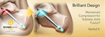
Intramedullary nailing is a type of surgery that repairs broken or diseased bones to keep them stable and help facilitate fusion. In this surgery, a permanent nail, i.e., an intramedullary nail or a rod is placed in the centre of the bone, so it would enable you to put weight on the bone. Typically this bone fixation procedure is carried out mostly in the Femur, Tibia, hip, and Humerus. Evolution of Hindfoot Fusion Intramedullary Nail Fixation During the 1 st generation of hindfoot fusion nailing surgery, there was no application of compression; instead, the nails were locked into the bone with screws. The 2 nd generation nail fixation applied external manual compression through the frame. 3 rd generation introduced internal compression by using an internal screw to press against the calcaneal screw. However with all these earlier generation nails, the bones are able to retain compression after the instruments are removed but would lose compression upon any bone resorption or s...

What are the Stages of Charcot foot?

Image
  “And miles to go before I sleep and miles to go before I sleep”! To walk a million miles in your life, you need your feet to support you in every step that you take, but Charcot foot or Charcot arthropathy can be a cause of concern. We, at MedShape Inc , provide effective surgical solutions to help address foot deformities and pain caused by Charcot arthropathy so that your feet support you in the way you want them to support you. What is Charcot foot? Charcot arthropathy or Charcot foot is a disease that affects soft tissues, joints and bones in the feet. From near-numbness to numbness in one or both feet, Charcot foot weakens the bones making them prone to fractures and dislocation. If Charcot foot remains untreated, it can further lead to rocker-bottom foot(when the shape of the foot changes in which the arch extends out and down),disability and amputation. What are the stages of Charcot foot? The 3 stages of Charcot foot are: ·       ...

What is Subtalar Fusion?

Image
Subtalar fusion is a type of surgical procedure performed to relieve pain and correct deformities in the hindfoot often affected by injury, arthritis, or genetic defect. Subtalar fusion involves fusing the calcaneus to the talus. The inferior tibiofibular joint is where the fibula meets the shin bone, and the subtalar joint is where the calcaneus (i.e. heel bone) meets the talus bone. Subtalar fusion is sometimes performed arthroscopically to relieve foot pain and reduce the amount of surgical exposure . Make sure to discuss with a reliable surgeon to know which method would be suitable for you. 1.       Open surgical procedure Open surgery for subtalar fusion is generally performed under general anesthetic and takes about ninety minutes. The surgeon will first make an incision on the outside of the ankle. Then, they will clean the joint surface and fix the joints together via one or two screws by implanting them through the back of the heel. The screws ...

Advanced Technology in Soft Tissue Fixation

Image
Soft Tissue Fixation Devices are designed for fixation of soft tissue to bone in tenodesis, ligament reconstruction and tendon transfer procedures. The design of the Eclipse™ Soft Tissue Anchor eliminates the hassles associated with other tenodesis devices. Those Tenodesis Devices such as interference screws can have issues such as rotating or lacerating the tendon. In the case of the Eclipse™ Soft Tissue Anchor , you can simply pull the trigger to watch it expand and lock the soft tissue to the bone .  There are a number of associated benefits including: Easy insertion The compressed profile of the Eclipse Sheath mates with soft tissue in the tunnel to provide easy insertion.  Non-rotational deployment The non-rotational deployment approach preserves the surgeon’s preferred soft tissue orientation.  Aids in facilitating healing The Eclipse effectively compresses the soft tissue in the bone tunnel that helps promote successful soft tissue-to-bone h...

What is Diabetic Charcot and how it can be Reversed?

Image
Diabetic Charcot Foot is a condition that affects the feet of people diagnosed with diabetes. It is also known as Charcot arthropathy . It mostly occurs in people that have lost the sensation or detected neuropathy. Charcot tends to simulate the joints, bones, delicate tissues of the ankle or foot. This ailment can make the bones turn weak that can break bones. Also, it can lead to the dislocation of the ankle or foot. It can damage the foot shape and can even direct to disability. The Charcot Ankle can lead to the deformation that spots unusual pressure on the outer side of the ankle.   The diabetic Charcot cannot be reversed because a human body cannot repair the damaged nerve tissues naturally. Still, you can try some practices to control the chances of increasing harm. Have control over blood sugar levels. Maintaining weight is essential to reduce the sensitivity of insulin. It increases the levels of blood sugar and can damage nerves. Consumption of high fiber and plant-...

All You Need to Know About the DynaNail Mini®Fusion System for Medial Column Fusion

Image
Charcot foot is a rare condition that affects people with peripheral neuropathy or who suffer from diabetes mellitus. The condition affects the joints, bones and soft tissues of the ankle or foot; whereby,the bones become brittle and prone to fractures. If not caught and treated in the early stages of development, patients suffer foot collapse and deformation. These deformities can lead to the development of pressure sores or ulcers in the ankle or foot. These open wounds can, in turn, become infected, eventually leading to amputation. Surgery is used as a last resort and is only recommended for patients with severe foot or ankle deformities and are at high risk of developing foot ulcers. The goals of the surgery are to stabilize the fractured bones and allow them to heal properly. The surgery involves realigning and stabilizing the bones to encourage healing in a position that allows the patient to weight-bear. In order to achieve healing and keep stable, continuous compression mu...

Different Reasons to Choose the Morphix® Suture Anchor Over Other Suture Anchors in Soft Tissue Repair Procedures

Image
 One of the most difficult aspects of orthopedic surgery is attaching soft tissues to bone. This aspect of tissue repair was previously left solely to the body’s natural healing mechanisms. This required the formation of adequate scar tissue. However, in order to achieve this, the limb or joint had to be immobilized for a long period. This ensured that the tissue was maintained in the correct position for the formation of scar tissue. Techniques for soft tissue attachment were later developed to improve soft tissue placement and fixation. These techniques included drilling bone tunnels with pull-through sutures or the use of fixation devices such as staples, screws, washers and tapered plugs. These fixation systems required larger access to the bone resulting in higher potential for damage during the tissue repair surgery. Because of their size and design, they often caused noticeable discomfort to patients and were also unable to provide adequate tension and stability in loadi...

Everything You Need to Know about DynaClip Forte Compression Staple Bone Fixation System

Image
Prior to the launch of Nitinol-based DynaClip® Bone Fixation System in 2019, plate and screws devices had predominantly been used in foot surgeries for bone fixation. Although Nitinol staples have proven effective for many foot and ankle bone fixation procedures, their application was limited due to their inability to withstand the mechanical demands of certain environments. The introduction of DynaClip as a bone fixation system offered a robust solution to maintain compression and the implant’s structural integrity long after surgery. Its design ensures the distribution of stress away from the corners of the implant thus preventing implant failure. MedShape Inc.’s launch of DynaClip Forte® compression staple builds on the 2-leg DynaClip bone staple. DynaClip Forte is a 4-leg foot staple with a low profile which allows the implant to rest on the bone. Patients don’t have to deal with irritation or complications arising from prominent or bulky hardware. DynaClip Forte’s four-leg de...